260 slides
High Throughput
One instrument supports both fluorescence and brightfield imaging, with fast switching, high-resolution brightfield imaging, multi-channel fluorescence, and up to 7-channel fluorescence imaging
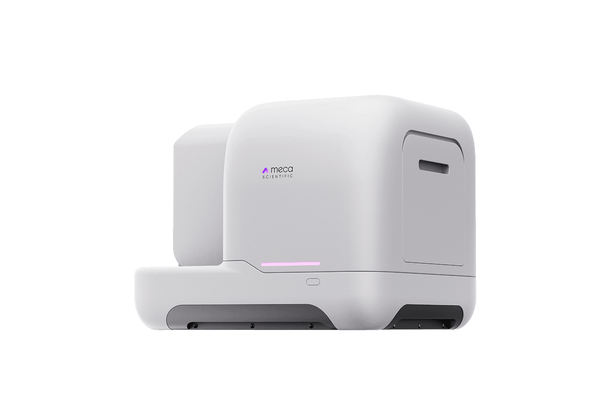
PanoFlow / High-throughput Brain Slice Analyzer
PanoFlow is a high-throughput brain slice analyzer built on PanoBrain, with higher throughput and a smoother research workflow experience
Apply for Trial260 slides
One instrument supports both fluorescence and brightfield imaging, with fast switching, high-resolution brightfield imaging, multi-channel fluorescence, and up to 7-channel fluorescence imaging
90 slides/h
The high-throughput brain slice analyzer can scan 90 slices within a 15 × 15 mm² area using 20X brightfield scanning
Progress Alerts
Large batch scanning can take time. The system automatically sends a message after scanning is complete, so you can track progress even when away from the instrument
Intuitive
The interface is clean and intuitive. Users can become familiar with the workflow in just 5 minutes and start slide scanning smoothly
No Breakage Risk
Slides move synchronously with the slide holder, effectively avoiding breakage risks that may occur when pushing individual slides
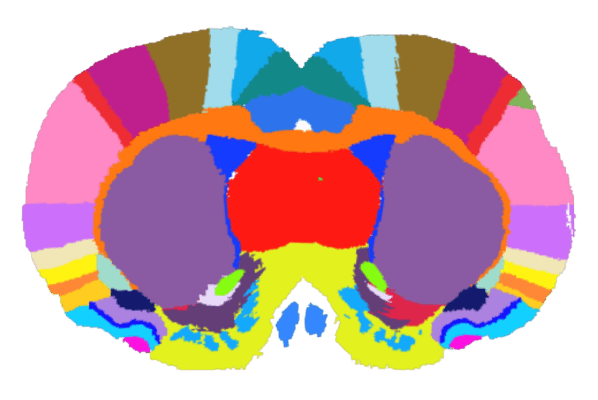
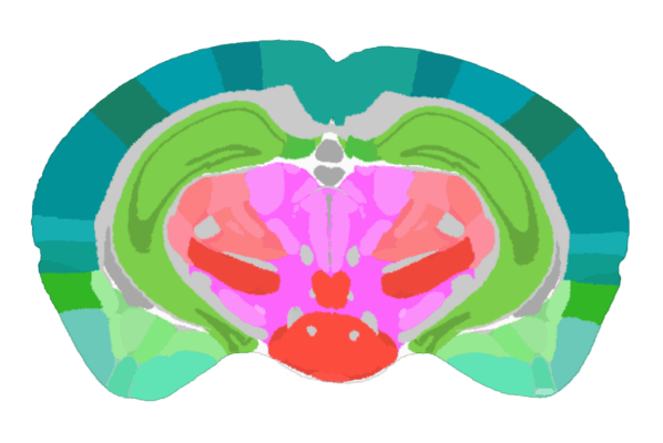
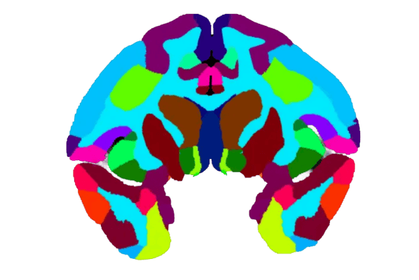

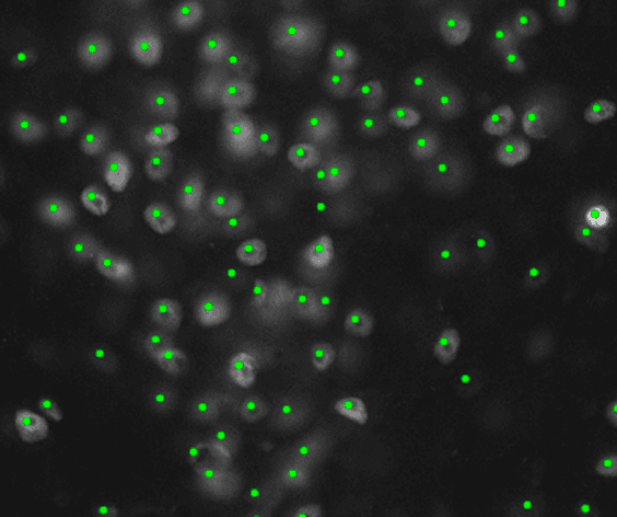
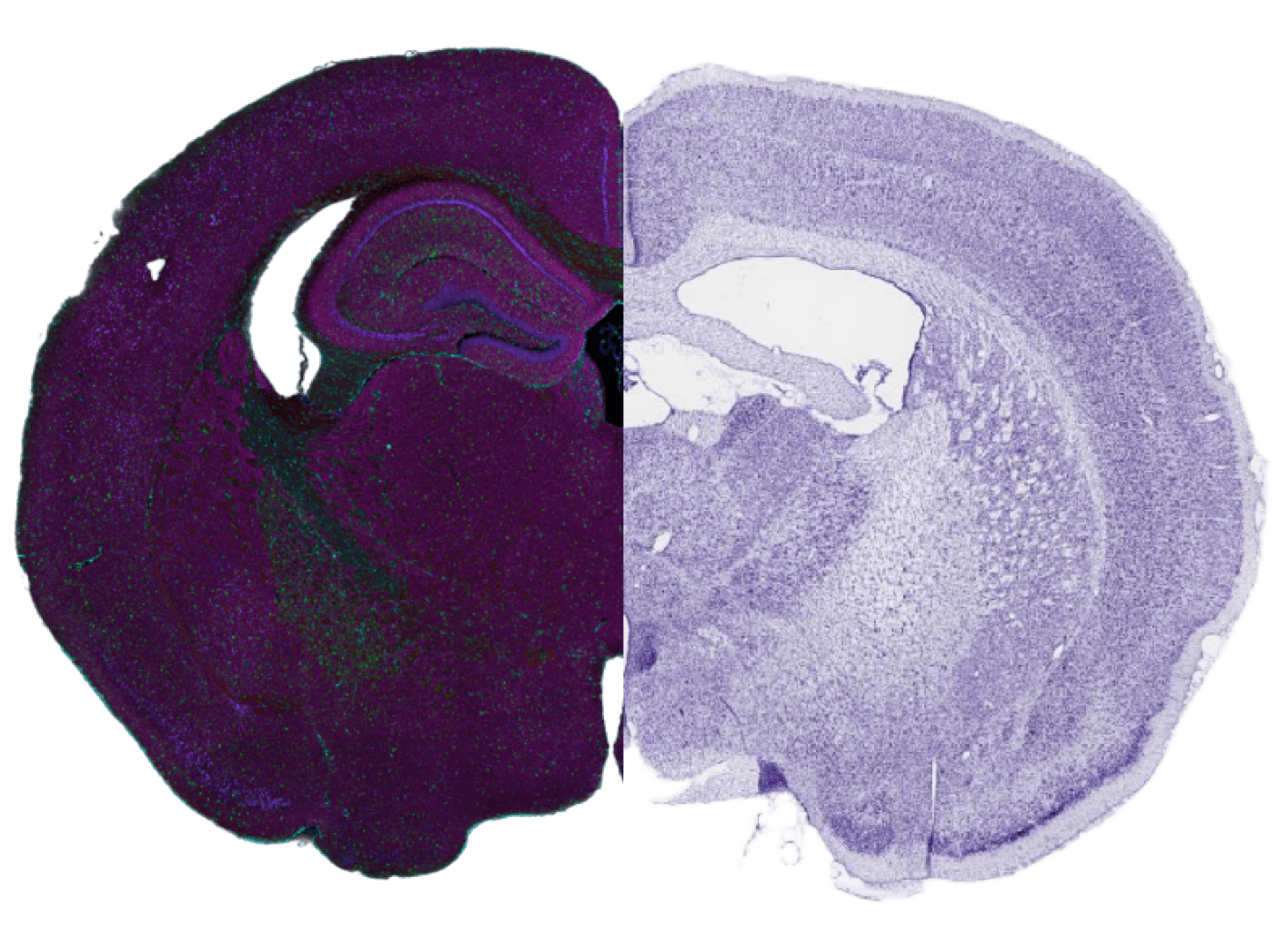
Supports automatic recognition of DAPI, c-Fos, neurons, glial cells, and other signals, and counts positive signal cells
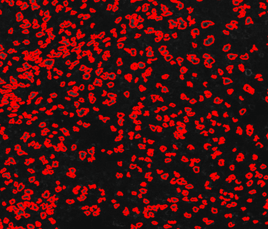
Supports automatic recognition of DAPI, c-Fos, neurons, glial cells, and other signals, with statistics for positive signal area and fluorescence intensity
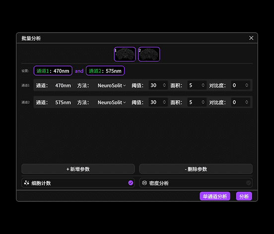
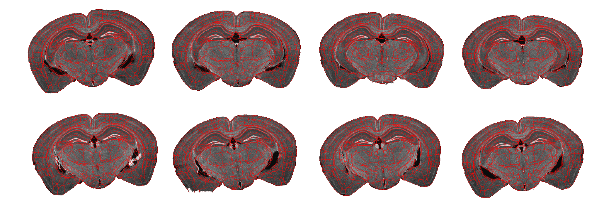
Supports batch analysis of multiple brain slices on the same slide, reducing repetitive manual work, improving analysis efficiency, and ensuring parameter consistency
One instrument supports both fluorescence and brightfield imaging modes, with fast switching between the two
Supports 7-channel fluorescence imaging with general channel configurations for common applications, plus customizable filters to reduce crosstalk concerns
Z-Stack scanning and extended depth of field fuse images from different focal planes in real time, producing fully focused images with clear layered details. Clear images can also be selectively extracted from the image sequence and stacked, reducing multi-layer fusion blur caused by defocus
Uniform illumination and global fluorescence background correction achieve up to 99% fluorescence background uniformity for seamless panoramic images
Strobe illumination provides millisecond-level high instantaneous power only during camera exposure, reducing fluorescence quenching. Scanned samples can still be used for confocal imaging
Custom modules are available for multi-channel fluorescence, large slide holders, sCMOS cameras, and throughput expansion
Designed for daily batch scanning in medium-throughput laboratories, balancing efficiency and footprint
For more frequent serial section tasks, helping teams steadily improve sample processing efficiency
A higher-throughput configuration for large-scale brain slice scanning and long unattended acquisition sessions
Custom modules are available for multi-channel fluorescence, large slide holders, sCMOS cameras, and throughput expansion
Move high-throughput brain slice scanning, analysis, and result delivery into a steadier automated workflow
Apply for Trial