Efficient Quenching
Rapidly and thoroughly removes fluorescence signal to prevent channel crosstalk, ensuring a pristine background for every imaging round and improving data reliability
FluoQ / Multiplex Unlocking
FluoQ fluorescence quenching system efficiently eliminates prior-round fluorescence signal and autofluorescence, unlocking the full picture of complex biological structures
Apply for TrialRapidly and thoroughly removes fluorescence signal to prevent channel crosstalk, ensuring a pristine background for every imaging round and improving data reliability
Breaks free from spectral channel limits with multi-round staining cycles, enabling hyperplex fluorescence labeling and multi-dimensional observation of complex biology
Compatible with paraffin sections, frozen sections, cell crawls, thick tissue sections, and more — covering pathology, immunology, and broader research scenarios
Residual signal is the hardest variable to control in multi-round imaging. FluoQ delivers complete quenching after each round — autofluorescence included — so the next antibody incubation begins on a genuinely clean background
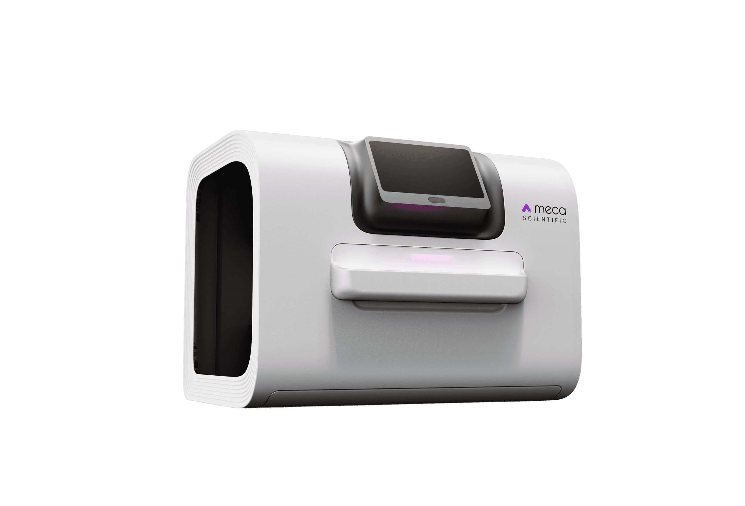
Capture high-quality fluorescence images of the current round's targets using PanoCube, PanoFlow, or other Meca imaging instruments — ensuring data completeness
Load the sample into FluoQ, add the matched quenching kit. The system runs the automated quenching protocol, thoroughly clearing the round's fluorescence signal
With a pristine background, begin the next round of fluorescent antibody labeling on the same sample — no residual interference, sharper signal interpretation
Repeat the stain–image–quench cycle to accumulate dozens of markers on a single sample, unlocking the full picture of complex biological structures
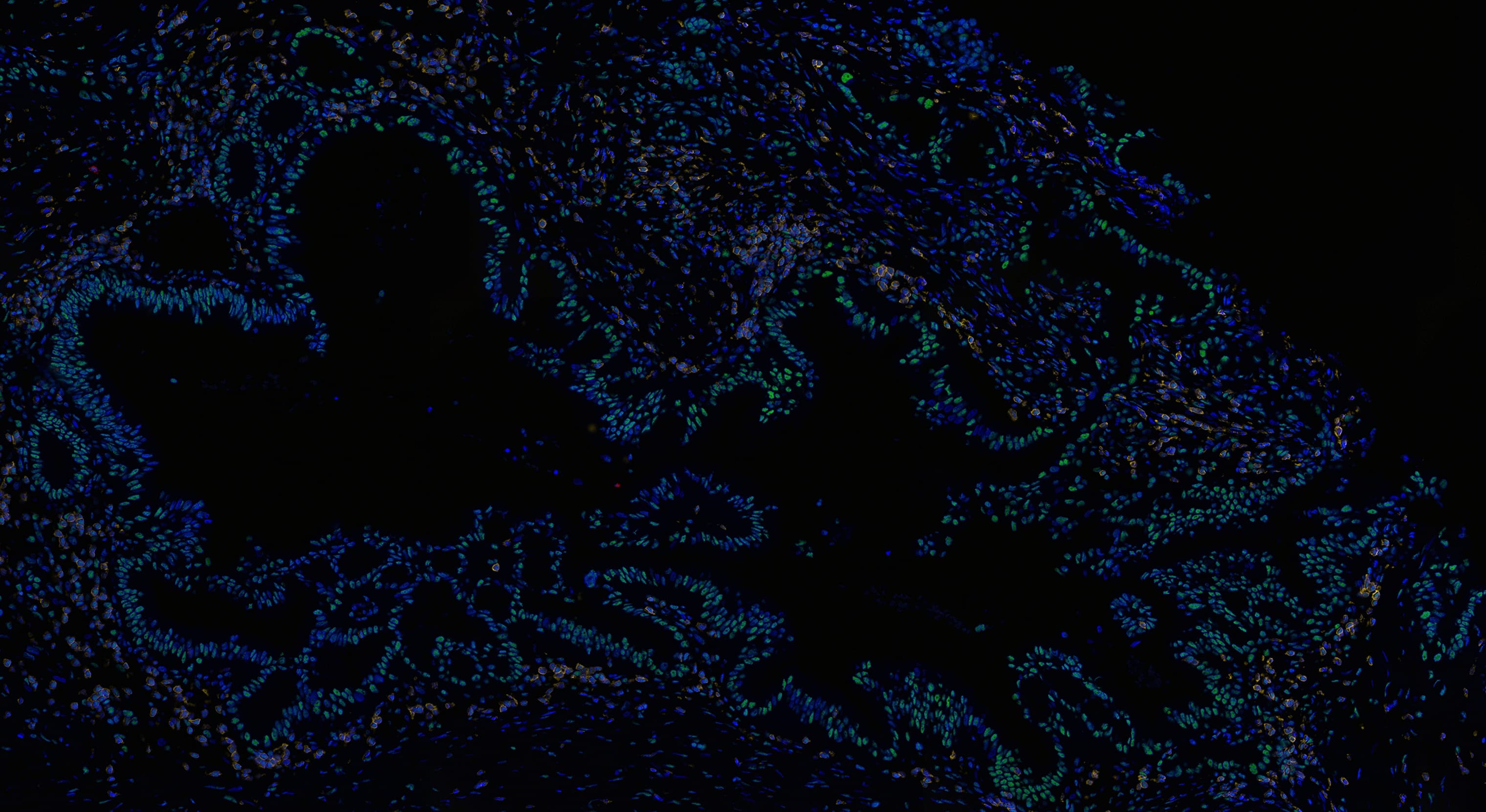
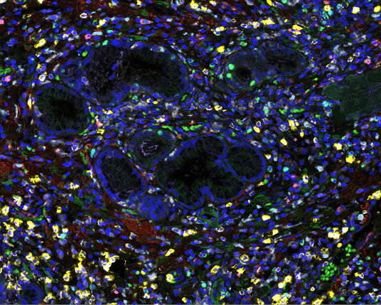
Round 1 stained section: CREPT, CD3, CD20
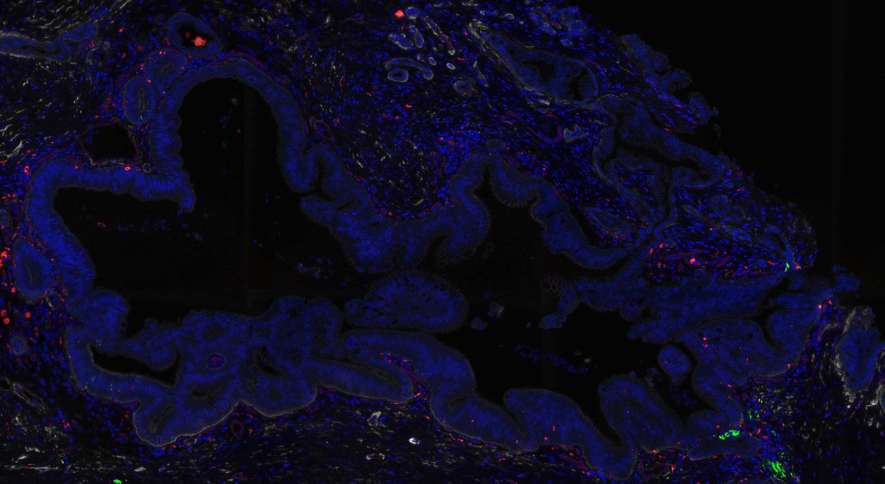
Round 2 stained section: CD56, CD68, SMA, CK19, CD31
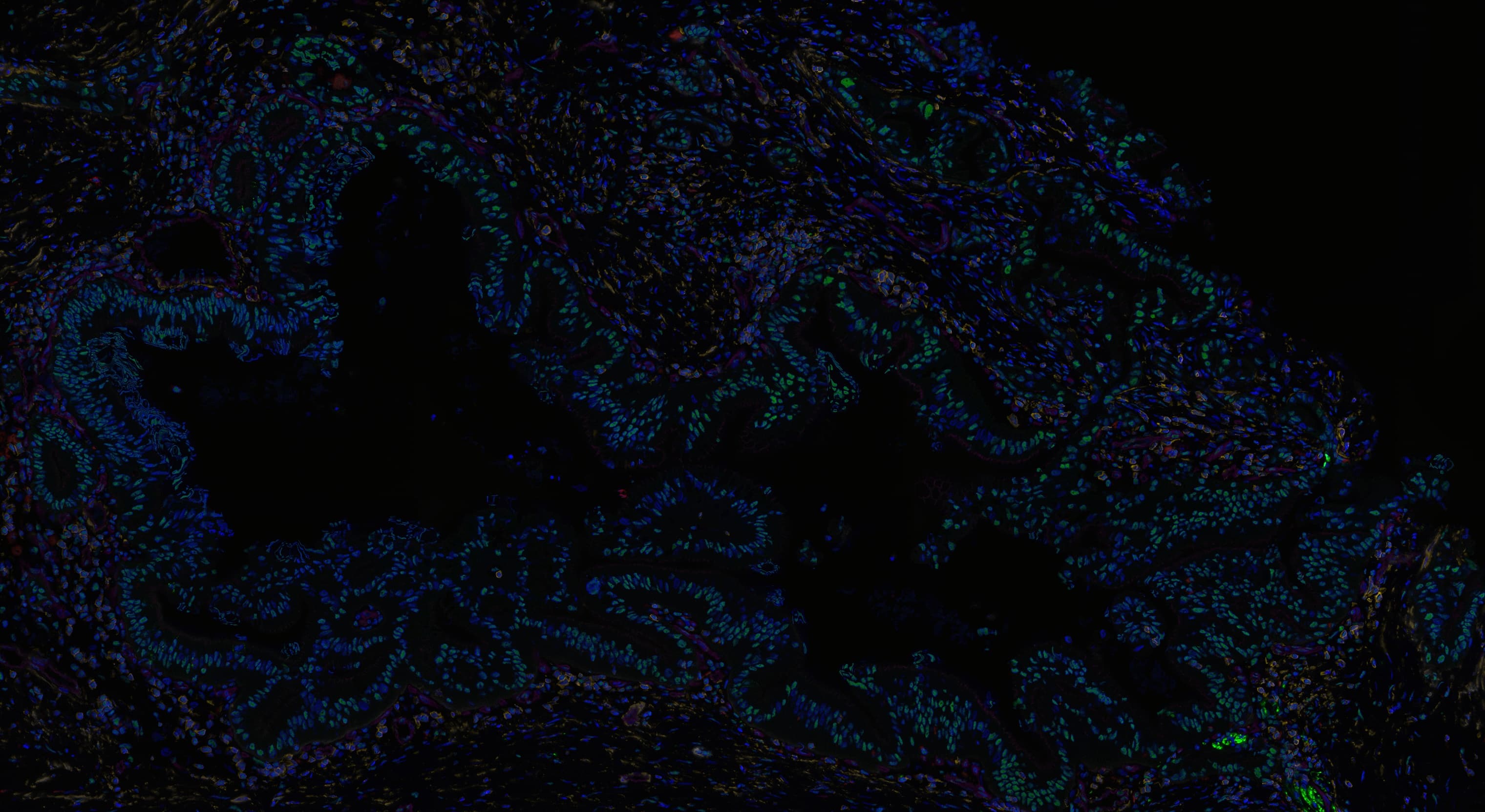
Section overlay: registered by the DAPI channel to generate a hyperplex image
The cap on fluorescent labels has never been the antibody — it's bleaching. FluoQ makes the same section reusable round after round. Each cycle lets you reincubate with a fresh antibody panel; markers stack linearly with cycles. Experiments that once needed multiple sections now fit on one
Continuously label a dozen-plus protein markers on a single tissue section, achieving information density beyond conventional multicolor while preserving full spatial context
Pair with platforms like Visium and Xenium to add a protein-level spatial layer to transcriptomic data; images plug directly into co-registration workflows
Biopsies, surgical margins, and other irreplaceable samples often yield only one section. FluoQ's low-damage chemistry lets a single section carry far more dimensions of measurement
High autofluorescence has long been the bottleneck for multicolor imaging in neural tissue. FluoQ's built-in autofluorescence suppression markedly improves SNR in brain sections
Simultaneously assess target expression, immune infiltration, and tissue architecture on a single section — a complete spatial evidence chain for candidate-drug mechanisms
Automated workflow makes multi-round imaging of hundred-case TMAs operationally feasible, with standardized acquisition that reduces batch variability for downstream analysis
Start from your experimental needs — let's discuss what multiplex imaging can unlock for you
Apply for Trial